


What Is A Stent For Kidney Stones? Painless Removal
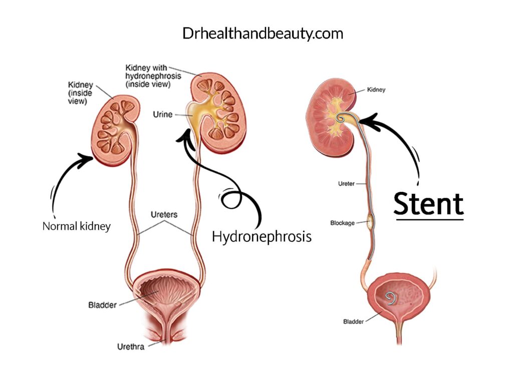
Ureters are tubes in the body that drain urine from the kidneys to the bladder. If one of your ureters is blocked, urine does not drain properly. When this happens, the kidney fills with urine and becomes swollen, called hydronephrosis. This can be caused by tumor pressure on the ureter, kidney stones, or scar tissue. It can be damaged if the kidney is blocked for a long time. If both of your ureters are blocked, this weakens your kidneys and puts you at risk of kidney failure. Obstructed kidney(s) requires the placement of a ureteral stent.
A stent for kidney stones is a thin tube placed in your ureter to help drain urine from the kidney. One end of the tube is inside the kidney, and the other is in the bladder.
Table of Contents
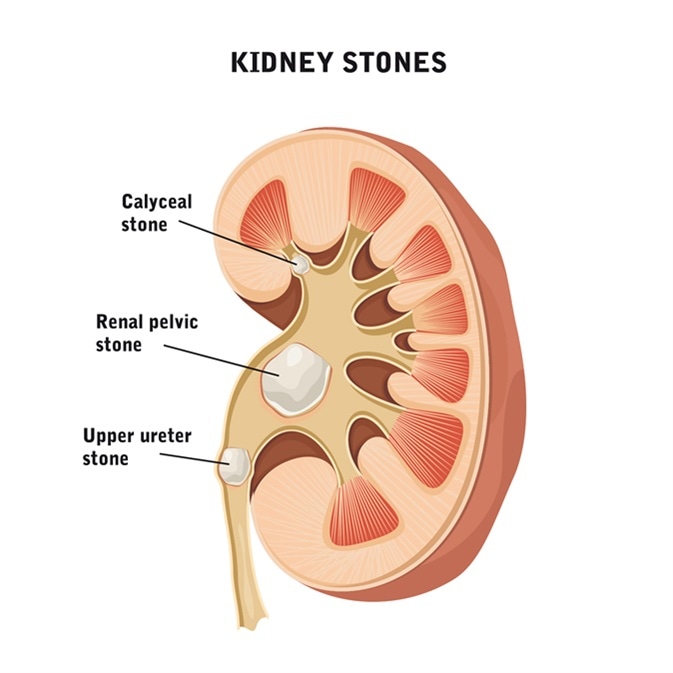
A ureteral stone is a kidney stone. A kidney stone has moved from the kidney to another part of the urinary tract.
The ureter is a tube that connects the kidney to the bladder. Its width is almost the size of a small vein, which is the most common place for kidney stones to cause pain, and depending on its size and location, it may be excruciating. If this stone is not removed, it causes untreatable pain or vomiting; sometimes, it may be accompanied by a fever or infection that should be referred to a doctor. (Urinary tract stones are relatively common).
stent for kidney stones
The tube that takes the urine from the kidney to the bladder is called the ureter tube; the blockage in the ureter tube can block the urine flow and cause swelling and damage to the kidneys; nephrostomy or stent for kidney stones are the ways. That the doctor It is used to make urine flow again.
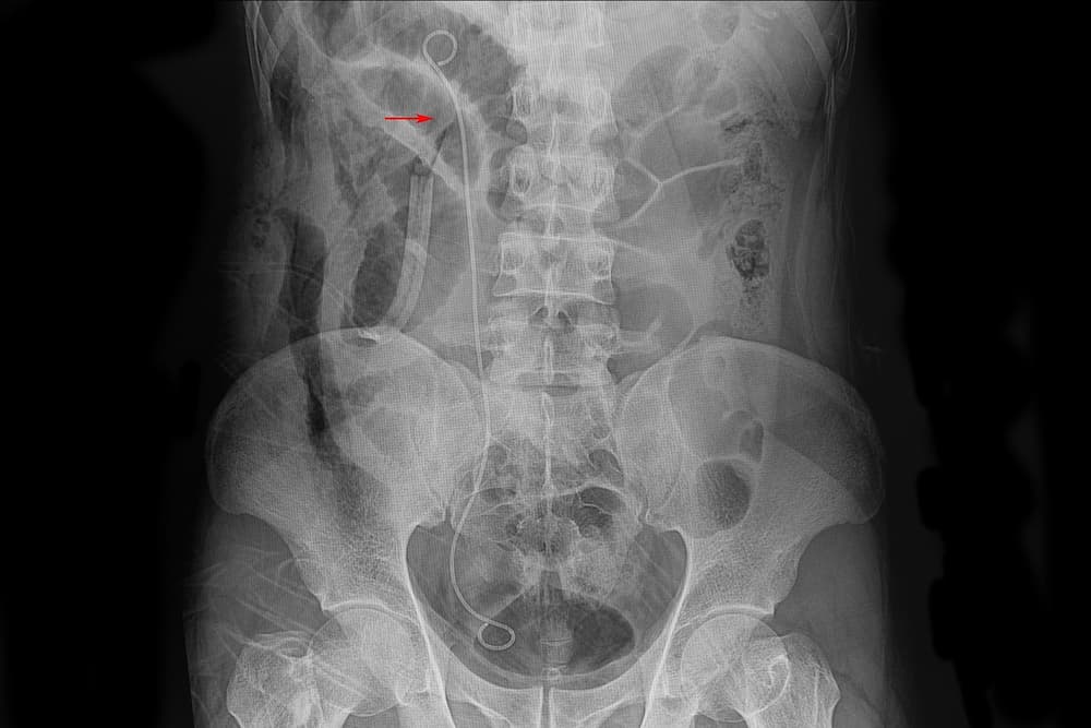
A stent for kidney stones is a tube whose two ends are twisted and ring-like; this device is transferred to the ureter tube by guiding the imaging device, which we will discuss later.
How are ureteral stones formed?
Kidney stones are a collection of crystals that usually form in the kidneys. These masses can form and move anywhere in the urinary tract, which includes the ureter, urethra, and bladder.
A ureteral stone is a kidney stone inside one of the ureters (tubes that connect the kidneys to the bladder). Stones are formed in the kidney and enter the ureter with urine.
– Sometimes, these stones are tiny. In this case, the stones may pass through your ureter, enter your bladder, and eventually pass out of your body when urinating. Sometimes the stone may be too big to pass and get stuck in the ureter, blocking the flow of urine and being very painful.
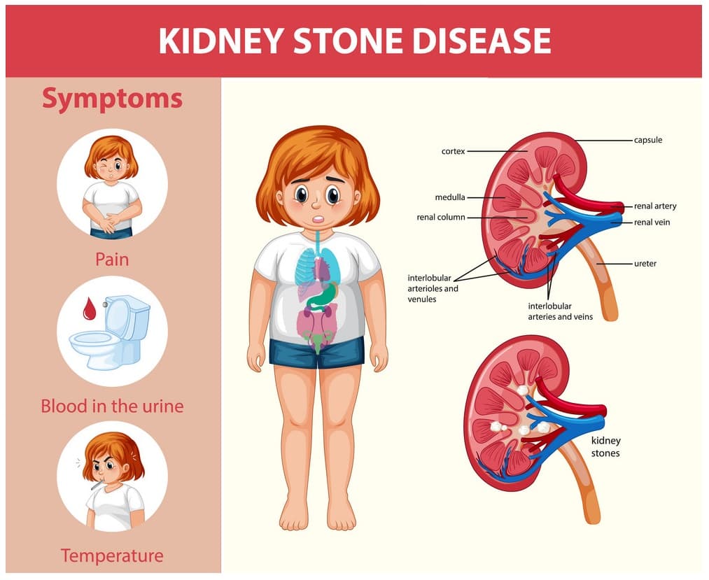
Symptoms of having ureteral stones
The most common symptom of kidney stones or ureters is pain.
You may feel pain in the lower abdomen or side. This pain can be mild and dull or annoying. The pain may also come and go and spread to other areas.
Other possible symptoms include:
- Pain or burning sensation when urinating
- Seeing blood in the urine
- Frequent urination
- nausea and vomiting
- Fever
If you experience any of these symptoms, you should see a urologist.
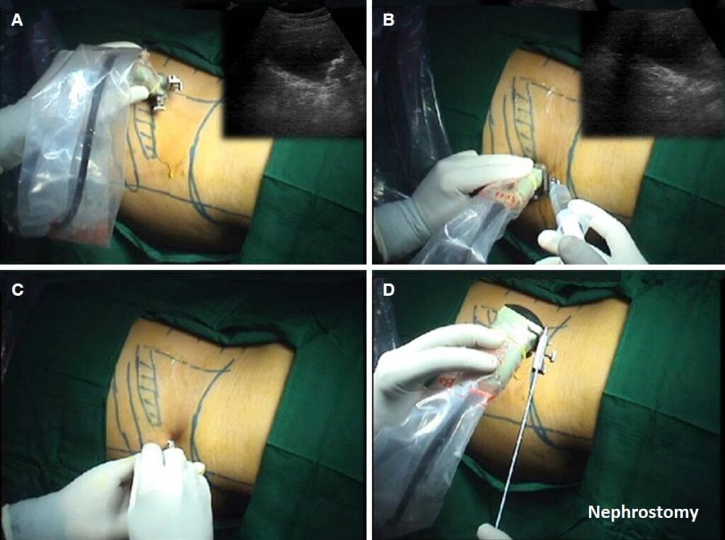
nephrostomy
If it is impossible to place a stent for kidney stones for the patient, the nephrostomy method is used. Nephrostomy is a procedure during which a tube is passed through the skin on the patient’s back to the kidney and connected to an external drainage bag.
stent for kidney stones and nephrostomy
As mentioned, urine is usually transferred from the kidney to the bladder through narrow tubes called the ureter; in some cases, the presence of kidney stones, infection, tumors, or blood clots obstruct the ureter, and the transfer of urine is difficult, which is due to Those secondary problems are caused by kidney diseases. The ureter, which helps to remove this blockage, means placing a narrow tube in the ureter, done with the ultrasound machine’s guidance.
Cancers that block the ureter!
In addition to infections, blood clots, and kidney stones, cancers that affect the patient in the abdomen and pelvis can also cause ureter obstruction. Among these cancers, the following can be mentioned:
- Bladder Cancer
- Colon cancer
- the rectum
- cervix
- womb
- ovary
- and prostate cancer
If, for any reason, it is not possible to place a stent, a nephrostomy procedure is performed.
Methods of diagnosing ureteral stones
If you have pain in your lower abdomen or notice blood in your urine, your doctor may order a kidney scan to detect stones.
Two of the most common imaging tests for stones are:
- Computed tomography (CT) scan: A CT scan is usually the best option for detecting stones in the urinary tract. It uses rotating X-ray machines to create cross-sectional images of the inside of the abdomen and pelvis.
- Ultrasound: Ultrasound does not use any radiation. This method uses high-frequency sound waves to produce images of the inside of your body.
These tests help your doctor determine the size and location of your stone. Knowing where and how big the stone is helps the doctor choose the right treatment plan.

Treatment of ureteral stones
“Research shows that many urinary stones go away without treatment.”
You may experience some pain as you pass them, but as long as you do not have a fever or infection, you may not need to do anything other than drink lots of water to pass the stones.
Small stones usually pass more quickly. The size of the stone is essential. Some stones, especially larger stones, get stuck in the ureter, the narrowest tube in your urinary tract. This can cause severe pain and increase the risk of infection.
If you have a bigger or broader stone, it is unlikely to pass independently; you must see a urologist.
Your doctor may recommend one of the following methods to remove a ureteral stone that is too large to pass on its own:
- Stent placement for kidney stones: A small, soft plastic tube is passed into the ureter around the stone, allowing urine to bypass the stone. This temporary solution is a surgical procedure that is performed under anesthesia.

- Nephrostomy tube placement: An internal radiologist can temporarily relieve pain by inserting this tube directly through the back into the kidney using only sedation and a combination of ultrasound and X-rays. It is usually used in case of fever or infection with urinary obstruction.
- Shock wave lithotripsy (ESWL): This procedure uses focused shock waves to break stones into smaller pieces, passing through the rest of your urinary tract and passing out of your body without additional assistance.
- Urethroscopy: Your urologist inserts a thin tube with a scope into your urethra and up into your ureter. When the doctor sees the stones, he removes them or divides them into smaller pieces with a laser so that they are expelled by themselves. This procedure may be performed by placing a ureteral stent to dilate the ureter in the weeks prior to ureteroscopy passively.
- Percutaneous Nephrolithotomy: This procedure is usually used if you have a very large or abnormal kidney stone. The doctor will make a small incision in your back and remove the stone through the incision with a nephroscope. Although this is a minimally invasive procedure, you will need general anesthesia.
- Drug therapy: This treatment involves using alpha-blocker drugs to help expel stones. Alpha-blockers help lower blood pressure, which can be effective for clearing smaller stones, but also carries a risk of adverse events. Has it?

How is stenting for kidney stones and nephrostomy performed?
In cases where it is possible to place a stent for kidney stones through the bladder and the patient’s health conditions allow it, urologists direct the device to the kidney in this way, but if this is not possible, interventional radiologists (International Radiologist) This procedure is performed on the skin of the patient, this procedure is mainly performed on an outpatient basis unless in some instances the doctor feels the need to hospitalize the patient.
Before placing a stent for kidney stones, it may be necessary to perform diagnostic tests such as ultrasound, MRI, or CT (Computerized Tomography), as well as possibly taking some antibiotics, painkillers, or anti-nausea drugs by the doctor. Recommended.
Complications of the stent for kidney stones
Some of the complications that stent for kidney stones can cause are listed here:
- Bleeding in the urine
- Dysuria or pain and burning during urination
- Frequent urination
- Bladder spasm or blockage
Sometimes, there may be no typical symptoms, and you should see a doctor when you see these conditions.
- Heavy bleeding in the urine or dark urine cannot be resolved by increasing fluid intake.
- A high fever is 101 degrees Fahrenheit (38.3 degrees Celsius) or higher.
- The presence of severe pain that is not relieved by painkillers.
- Pain in both sides of the abdomen or kidney.
- Bad smell in urine.
- Burning sensation when urinating.
- cloudy urine
Preparation for a stent for kidney stones
After performing the initial stages of diagnosis, including MRI, CT, ultrasound, and blood tests, the doctor will inform you of the time for the stent or nephrostomy. When this procedure is to be performed, the patient must stop eating at midnight. Refrain; if you need medicine, drink it with a small amount of water.
This procedure is usually performed outpatient, and the doctor will tell you if hospitalization is necessary.
After placing a stent for kidney stones
You cannot drive after the procedure, so choose someone to drive you home when you are done.
After the operation of the stent for kidney stones, some things must be observed.
In the twenty-four hours after the operation, water consumption is significant, and you should drink more water than usual, between 8 and 10 glasses of water. The patient should not lift heavy objects during this period (more than 6.8 kg) or Any activity that uses the abdominal muscles. After 24 hours, the person can resume his normal activities.
After placing a stent for kidney stones, the patient may experience conditions such as:
- Frequent urination
- Strong and sudden urge to urinate
- Pelvic and abdominal pain
Moreover, these symptoms usually disappear with time, and the doctor and nurse will explain the situation to you after the operation.
Similarly, in some cases, the patient may see blood in his urine, which may remain as long as the stent is in place, which can be resolved by drinking more water.
Stent replacement
After 3 to 6 months, the stent must be replaced, and the doctor or nurse will explain how long it will take.
Leaving a ureteral stent for kidney stones in place may lead to ureteral blockage, kidney stones, and infection.
Conclusion
A stent for kidney stones or double-J or ureteral stent is a thin, flexible tube that keeps the ureter open. The ureter is a part of the urinary system, a long and thin tube carrying urine from the kidneys to the bladder. The doctor places the ureteral stent inside this tube-like passage. One end of this stent is placed inside the renal pelvis, and the other reaches the bladder after passing through the ureter. Because the two sides of this tube look like the letter J, it is also called “double J.”
A stent for kidney stones is usually placed during kidney stone surgery. Of course, the use of this stent is not limited to kidney stone surgery; the function of this stent is to keep the ureter open so that the stones or their remains can pass more comfortably without damaging the ureter. On the other hand, because kidney stone surgery may cause damage to the ureter, resulting in its swelling and narrowing, Double J is used to keep this duct open and pass urine and stones easily.
References:

Dr.sanaz bellucci
I am Sanaz Bellucci , a dentist. In this electronic magazine, I publish my collection of research and studies on various topics related to health and beauty that you may face with it.
You can also share your questions of interest with me so that I can give them complete and comprehensive answers. stay with me.
Share in :
Facebook
Twitter
LinkedIn
Reddit
Explore more

Dr.sanaz bellucci
September 12, 2023